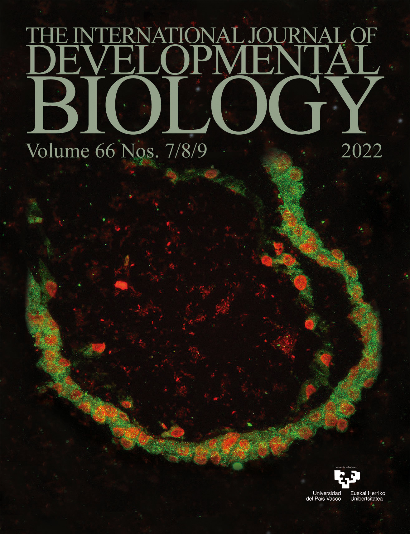
Cyclooxygenase-2 and the chamber of secrets: The developing heart of a chick, stained for the presence of previously unknown cyclooxygenase-2. (COX-2 in green and nucleus in red). For further details, see article by Parmar et al., pp. 373-381 in the current issue.
Regeneration and proliferation of cardiomyocytes and its microRNA regulatory mechanisms
Int. J. Dev. Biol. (2022) 66: 359-372
https://doi.org/10.1387/ijdb.220097yc
ABSTRACT
Myocardial regeneration is identified as a concept at histological level. The core content is to increase the number of cardiomyocytes (CMs), so as to maintain the steady state of CMs under pathological or physiological conditions and ensure the normal cardiac function. In this review, we discussed the relevant factors involved in the regeneration of CMs, generalized in mice, large mammals and human. During different development stages of mammalian hearts, CMs showed several controlling and growth modes on the physiological or pathological state: mitosis, hypertrophy, nuclear polyploidy and multinucleation, amitosis and etc. We also discussed the mechanisms of specific microRNAs implicated in the cardiac development, as well as disease-induced apoptosis in CMs and the process of re-entering cell cycle after injury. It is hoped that this review will contribute to a deeper understanding of therapeutic approaches for myocardial regeneration after injury.Cyclooxygenase-2 plays a crucial role during myocardial patterning of developing chick
Int. J. Dev. Biol. (2022) 66: 373-381
https://doi.org/10.1387/ijdb.220153sb
ABSTRACT
Cyclooxygenase-2 (COX-2), a member of the Cyclooxygenase family, initiates the biosynthesis of prostanoids that regulates various cellular functions. Our pilot attempt revealed that the administration of etoricoxib, an inhibitor specific for COX-2, induces abnormal looping in the chicken heart. The present study attempts to reveal the mechanistic details of etoricoxib-induced abnormal cardiac looping. The activity of COX-2 was inhibited by administering 3.5 μg of etoricoxib into the egg’s air cell on day zero of incubation. The gene and protein expression patterns of key mediators of heart development were then analyzed on day 2 (HH12) and day 3 (HH20). Reduced COX-2 activity altered the expressions of upstream regulators of organogenesis like Wnt11, BMP4, and SHH in the etoricoxib-exposed embryos. The observed expression shifts in the downstream regulators of myocardial patterning (MYOCD, HAND2, GATA4, GATA5, and GATA6) in the treated embryos corroborate the above results. In addition, the reduction in COX-2 activity hampered cardiomyocyte proliferation with a concomitant increase in the apoptosis rate. In conclusion, the collective effect of altered expression of signaling...The involvement of hormone-sensitive lipase in all-trans retinoic acid induced cleft palate
Int. J. Dev. Biol. (2022) 66: 383-389
https://doi.org/10.1387/ijdb.220137kz
ABSTRACT
Abnormally high concentrations of all-trans retinoic acid (atRA) induce cleft palate, which is accompanied by abnormal migration and proliferation of mouse embryonic palatal mesenchyme (MEPM) cells. Hormone-sensitive lipase (HSL) is involved in many embryonic development processes. The current study was designed to elucidate the mechanism of HSL in cleft palate induced by atRA. To establish a cleft palate model in Kunming mice, pregnant mice were administered atRA (70 mg/kg) by gavage at embryonic Day 10.5 (E10.5). Embryonic palates were obtained through the dissection of pregnant mice at E15.5. Hematoxylin and eosin (H&E) staining was used to evaluate growth changes in the palatal shelves. The levels of HSL in MEPM cells were detected by immunohistochemistry, quantitative real-time reverse transcription-polymerase chain reaction (qRT-PCR) and western blotting. RNAi was applied to construct vectors expressing HSL small interference RNAs (siRNAs). The vectors were transfected into MEPM cells. Cell proliferation and migration were evaluated by the cell counting kit-8 (CCK-8) assay and wound healing assay, respectively. The palatal shelves in the atRA group had separated at E15.5...Int. J. Dev. Biol. (2022) 66: 391-400
https://doi.org/10.1387/ijdb.220141db
ABSTRACT
Although histone methyltransferases are implicated in many key developmental processes, the contribution of individual chromatin modifiers in dental tissues is not well understood. Using single-cell RNA sequencing, we examined the expression profiles of the disruptor of telomeric silencing 1-like (Dot1L) gene in the postnatal day 5 mouse molar dental pulp. Dot1L is the only known enzyme that methylates histone 3 on lysine 79, a modification associated with gene expression. Our research revealed 15 distinct clusters representing different populations of mesenchymal stromal cells (MSCs), immune cells, pericytes, ameloblasts and endothelial cells. We documented heterogeneity in gene expression across different subpopulations of MSCs, a good indicator that these stromal progenitors undergo different phases of osteogenic differentiation. Interestingly, although Dot1L was broadly expressed across all cell clusters within the molar dental pulp, our analyses indicated specific enrichment of Dot1L within two clusters of MSCs, as well as cell clusters characterized as ameloblasts and endothelial cells. Moreover, we detected Dot1L co-expression with...