Edited by: Nikolas Zagris
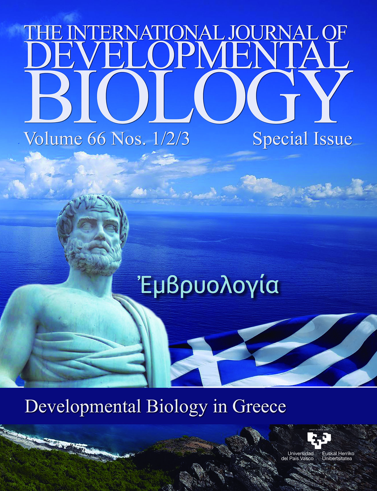
Ἐμβρυολογία/Embryology originated with Aristotle, the ‘Father of Natural History’. His treatise Περίζῴωνγενέσεως/On the Generation of Animals is the first ‘great compendium of embryology’ ever written, and is ‘by far the most important in the history of embryology’. The introduction of molecular techniques being applied to the classical problems of embryonic development transformed classical embryology to a field of codes and regulatory circuits. Developmental Biology has flourished and is now a vibrant interdisciplinary field in present day Greece.
OPEN ACCESS
Aristotle (384-322 BC): the beginnings of Embryology
Int. J. Dev. Biol. (2022) 66: 5-8
https://doi.org/10.1387/ijdb.220040nz
ABSTRACT
Aristotle made important contributions to many fields-biology, physics, metaphysics, logic, ethics, rhetoric, psychology, aesthetics, poetry- that are now cultivated by specialized experts, but he never lost sight of the aim of unifying knowledge, of understanding the world as an organized whole. Aristotle was the first to combine wet, field biology with daring cosmological thinking. He is the father of natural history and the first embryologist known to history. Aristotle’s classic treatises History of Animals/ΠΕΕί Ζῴων ἱΣτΟρίαι, and On the Generation of Animals/ ΠΕΕί Ζῴων ΓΕνέΣΕως “enjoyed for more than fifteen hundred years an authority altogether without parallel”. Over the last four decades, the introduction of molecular techniques has gradually overturned the entire structure of the biological sciences. Biology, initially a science of inventory and classification in the hands of the 19th-century comparative naturalists, has become a science of codes and regulatory circuits....Birth and death of neurons in the developing and mature mammalian brain
Int. J. Dev. Biol. (2022) 66: 9-22
https://doi.org/10.1387/ijdb.210139id
ABSTRACT
Although neuron birth and death are two contradictory processes, they serve the same purpose of the formation of the brain. They coexist during brain development, when cytoarchitecture and synaptic contacts are progressively established. It is the highly programmed interplay between these two processes that results in the making of a mature, complex-wired, functional brain. Neurogenesis is the process that begins with the birth of naïve new neurons, which are gradually specified to their prospective cell fate, translocate through migratory streams to the brain area they are destined for, and terminally differentiate into mature neurons that integrate into neuronal networks with sophisticated functions. This is an ongoing process until adulthood, when it mediates brain neuroplasticity. Neuron death is the process through which the fine sculpting and modeling of the brain is achieved. It serves to adjust final neuron numbers, exerting quality control over neurons that birth has generated or overproduced. It additionally corrects early wiring and performs systems matching by negatively selecting neurons that fail to gain neurotransmitter-mediated neuronal activity or receive...Organoids: the third dimension of human brain development and disease
Int. J. Dev. Biol. (2022) 66: 23-33
https://doi.org/10.1387/ijdb.210158gk
ABSTRACT
Stem cell technologies have opened up new avenues in the study of human biology and disease. In particular, the advent of human embryonic stem cells followed by reprograming technologies for generation of induced pluripotent stem cells have instigated studies into modeling human brain development and disease by providing a means to simulate a human tissue otherwise completely or largely inaccessible to researchers. Brain development is a complex process achieved in a remarkably controlled spatial and temporal manner through coordinated cellular and molecular events. In vitro models aim to mimic these processes and recapitulate brain organogenesis. Initially, two‐dimensional neural cultures presented an innovative landmark for investigating human neuronal and, more recently, glial biology, as well as for modeling brain neurodevelopmental and neurodegenerative diseases. The establishment of three‐dimensional cultures in the form of brain organoids was an equally important milestone in the field. Brain organoids mimic more closely the in vivo tissue composition and architecture and are more physiologically relevant than monolayer cultures. They therefore...Cortical interneuron development: a role for small Rho GTPases
Int. J. Dev. Biol. (2022) 66: 35-42
https://doi.org/10.1387/ijdb.210186dk
ABSTRACT
GABAergic interneurons control cortical excitation/inhibition balance and are implicated in severe neurodevelopmental disorders. In contrast to the multiplicity of signals underlying the generation and migration of cortical interneurons, the intracellular proteins mediating the response to these cues are largely unknown. We have demonstrated the unique and diverse roles of the Rho GTPases Rac1 and 3 in cell cycle and morphology in transgenic animals where Rac1 and Rac1/3 were ablated specifically in cortical interneurons. In the Rac1 mutant, progenitors delay their cell cycle exit, probably due to a prolonged G1 phase resulting in a cortex with 50% reductions in interneurons and an imbalance of excitation/inhibition in cortical circuits. This disruption in GABAergic inhibition alters glutamatergic function in the adult cortex, which could be reversed by enhancement of GABAergic functions during an early postnatal period. Furthermore, this disruption disturbs neuronal synchronization in the adult cortex. In the double mutant, additional severe cytoskeletal defects result in an 80% interneuron decrease. Both lines die postnatally from epileptic seizures. We have made progress...OPEN ACCESS
The development of MGE-derived cortical interneurons: An Lhx6 tale
Int. J. Dev. Biol. (2022) 66: 43-49
https://doi.org/10.1387/ijdb.210185md
ABSTRACT
The cerebral cortex contains two main neuronal cell populations: the excitatory pyramidal neurons and the inhibitory interneurons, which constitute 20-30% of all cortical neurons. Cortical interneurons are characterized by a remarkable morphological, molecular and functional diversity. A swathe of research activity over the last 20 years has sought to determine how cortical interneurons acquire their mature cellular and functional features, and has identified a number of transcription factors that function at different stages of interneuron development. Here, we review all current knowledge concerning the multiple functions of the “master regulator” - LIM-Homeodomain transcription factor Lhx6 - a gene expressed in the medial ganglionic eminence of the basal telencephalon that controls the development of somatostatin and parvalbumin expressing interneurons.Heterogeneity of quiescent and active neural stem cells in the postnatal brain
Int. J. Dev. Biol. (2022) 66: 51-58
https://doi.org/10.1387/ijdb.220010ik
ABSTRACT
In the postnatal mammalian brain, neurogenic activity is retained in anatomically restricted areas, driven by pools of Neural Stem Cells (NSCs). These cells and their progeny have been studied intensively as potential targets for regenerative treatments, aiming at either their in situ manipulation or their use as sources of cells for transplantation-based strategies. Although their full identity, heterogeneity and differentiation potential remain elusive, due to the absence of specific cell-type markers, our knowledge of their properties is constantly expanding. Here, we focus on the NSC niche that is located at the Subependymal Zone (SEZ/ also known as Subventricular Zone) of the lateral ventricles of the brain. We review, summarize and explain the different faces of the NSC, as they have been described, using a wide range of experimental approaches, over a time-frame of three decades: the primitive, definitive, quiescent or activated NSC. We also review the growing evidence of the existence of latent NSCs outside of niches, in the brain parenchyma, that constitute promising new therapeutic targets, complemented by the novel technologies of in vivo cell...Developmental and regenerative biology of cardiomyocytes
Int. J. Dev. Biol. (2022) 66: 59-75
https://doi.org/10.1387/ijdb.210159kh
ABSTRACT
Current progress and challenges in understanding the molecular and cellular mechanisms of cardiomyocyte embryonic development and regeneration are reviewed in our present work. Three major topics are critically discussed: how do cardiomyocytes form in the embryo? What is the adult origin of the cells that regenerate cardiomyocytes in animal models with adult heart regeneration capabilities? Can the promise of therapeutic cardiomyocyte regeneration be realized in humans? In the first topic, we highlight current advances in understanding the developmental biology of cardiomyocytes, with emphasis on the regulative capabilities of the early embryo during specification and allocation of the cardiomyoblasts that produce the primordial heart. We place further emphasis on trabecular cardiomyocyte development from late cardiomyoblasts, neural crest cells and primordial cardiomyocytes, and their critical role in the clonal growth of the compact/septal and cortical cardiomyocyte layers in the mammalian embryo and adult zebrafish, respectively. In the second topic, we focus on the re-activation of the cortical or trabecular compaction programs as hallmarks of cardiomyocyte regenerative cells...OPEN ACCESS
Int. J. Dev. Biol. (2022) 66: 77-83
https://doi.org/10.1387/ijdb.210167pk
ABSTRACT
Vascular Endothelial cadherin, a type II classical cadherin, is the major cadherin molecule participating in homotypic cell-cell adhesion structures between endothelial cells. It associates with cytoplasmic and membrane cytoskeletal elements to form endothelial adherens junctions (AJs), pivotal in regulating endothelial barrier function in the adult. VE-cadherin-mediated AJs are also involved in signaling via direct or indirect associations with receptors. The generation of mutant animals, especially mice and zebrafish, revealed many details concerning the role of VE-cadherin-mediated AJs in cardiovascular development. In general, VE-cadherin knockout (KO) in mice is embryonic lethal due to severe cardiovascular defects, and major signaling pathways as well as vascular formation cues were discovered in developing endothelium. However, there is little information regarding AJs formation and their components in cardiovascular progenitors. We have characterized in detail the activation pattern of mouse VE-cadherin promoter (Pvec) in a mouse embryonic stem cells (ESCs) differentiation system in vitro. Surprisingly, we found that it is activated transiently...Promyelocytic leukemia protein (PML) and stem cells: from cancer to pluripotency
Int. J. Dev. Biol. (2022) 66: 85-95
https://doi.org/10.1387/ijdb.210154av
ABSTRACT
The promyelocytic leukemia protein (PML) is the core organizer of cognate nuclear bodies (PML-NBs). Through physical interaction or modification of diverse protein clients, PML-NBs regulate a multitude of - often antithetical- biological processes such as antiviral and stress response, inhibition of cell proliferation and autophagy, and promotion of apoptosis or senescence. Although PML was originally recognized as a tumor-suppressive factor, more recent studies have revealed a “double-faced” agent role for PML. Indeed, PML displayed tumor cell pro-survival and pro-migratory functions via inhibition of migration suppressing molecules or promotion of transforming growth factor beta (TGF-β) mediated Epithelial-Mesenchymal Transition (EMT) that may promote cancer cell dissemination. In this line, PML was found to correlate with poor patient prognosis in distinct tumor contexts. Furthermore, in the last decade, a number of publications have implicated PML in the physiology of normal or cancer stem cells (CSCs). Promyelocytic leukemia protein activates fatty acid oxidation (FAO), a metabolic mechanism required for the asymmetric divisions and maintenance of hematopoietic stem...Exosomes and the extracellular matrix: a dynamic interplay in cancer progression
Int. J. Dev. Biol. (2022) 66: 97-102
https://doi.org/10.1387/ijdb.210120nk
ABSTRACT
Exosomes are a subtype of extracellular vesicles (EVs) composed of a lipid bilayer, which carry various cargoes such as nucleic acids, proteins, and bioactive lipids. Cancer cells release exosomes to promote cell communication and interaction with the extracellular matrix (ECM). ECM regulates the secretion and uptake of exosomes. Moreover, the cargo of exosomes can control ECM remodeling, thus affecting cancer progression. Aside from the rearrangement of ECM, exosomal cargo also modulates different signaling pathways that maintain homeostasis and play a major role in tumor growth and immune evasion in the tumor microenvironment (TME). Exosomes are now widely recognized as circulating biomarkers for diagnosis and prognosis. Their role in cancer initiation, progression, and chemoresistance is becoming increasingly clear from preclinical and clinical investigations, thereby gaining interest for their potential use as cancer diagnostics tools, but also for the development of future innovative cancer therapeutics. In this mini review we outline and discuss the correlation between exosomes, TME and cancer progression, while focusing on the potential role of exosomes as diagnostic and...Vascular cell-matrix adhesion in development and cancer
Int. J. Dev. Biol. (2022) 66: 103-113
https://doi.org/10.1387/ijdb.210204vk
ABSTRACT
The development and homeostasis of vertebrate organisms depend on the “tree of life”, in other words, the intricate network of vascular tubes composed of endothelial cells attached to the basement membrane and surrounded by perivascular cells. Although many studies have revealed the fundamental role of cytokines, growth factors and Notch signalling in vascular morphogenesis, we still lack sufficient understanding of the molecular mechanisms controlling the various steps of the angiogenic processes. Emerging data highlight that cell adhesions are key players in vascular morphogenesis. In this review, we focus on endothelial cells and we present the current state of knowledge regarding the role of cell-matrix adhesions in developmental and tumour angiogenesis, attained mainly from genetic studies and animal models.On the role of pleiotrophin and its receptors in development and angiogenesis
Int. J. Dev. Biol. (2022) 66: 115-124
https://doi.org/10.1387/ijdb.210122ep
ABSTRACT
The secreted growth factor pleiotrophin (PTN) is expressed in all species and is evolutionarily highly conserved, suggesting that it plays a significant role in the regulation of important processes. The observation that it is highly expressed at early stages during development and in embryonic progenitor cells highlights a potentially important contribution to development. There is ample evidence of the role of PTN in the development of the nervous system and hematopoiesis, some, albeit inconclusive, evidence of its role in the skeletomuscular system, and limited evidence of its role in the development of other organs. Studies on its role in the cardiovascular system and angiogenesis suggest that PTN has a significant regulatory effect by acting on endothelial cells, while its role in the functions of smooth or cardiac muscle cells has not been studied. This review highlights what is known to date regarding the role of PTN in the development of various organs and in angiogenesis. Wherever possible, evidence on the crosstalk between the receptors that mediate PTN’s functions is also quoted, highlighting the complex regulatory pathways that affect development and angiogenesis.Application of developmental principles for spinal cord repair after injury
Int. J. Dev. Biol. (2022) 66: 125-135
https://doi.org/10.1387/ijdb.210110fp
ABSTRACT
The superiority of the mammalian central nervous system (CNS) compared with other vertebrates does not involve an advanced capacity for regeneration, and any insult results in irreversible functional loss. Spinal cord injury (SCI) is one example of CNS trauma affecting thousands of individuals, mostly young, each year. Despite enormous progress in our comprehension of the molecular and cellular mechanisms underlying the pathophysiology after SCI, also providing targets for therapeutic interventions, no efficient therapy exists as yet, emphasizing the need for further research. A breadth of studies have demonstrated that, after SCI, principles of development come into play either to promote or to prohibit spontaneous regeneration, and their appropriate manipulation has the potential to contribute towards functional recovery. In this overview, some of the most recent and important studies are discussed.These offer explicitly novel input from the field of development to the field of CNS repair regarding the modification of the inhibitory environment of the injured spinal cord – mainly referring to the glial scar – the activation of endogenous cell populations such as ependymal...OPEN ACCESS
Int. J. Dev. Biol. (2022) 66: 137-154
https://doi.org/10.1387/ijdb.210108sa
ABSTRACT
This review is an update with regard to the efforts to develop liposomal carriers for growth factor delivery. It is well known that growth factors have the potential to enhance/accelerate tissue regeneration; however, their poor stability, which results in rapid loss of their activity, together with their rapid clearance from defected tissues (when applied as free molecules) is a serious drawback for their use; their highly hydrophilic nature and low capability to permeate through biological barriers (cell membranes) are additional factors that limit their applicability. In recent years, the advantages of liposomal drug delivery systems have motivated efforts to deliver growth factors (GFs) in liposomal form. Herein, after briefly introducing the basic structural characteristics of liposome types and their advantages when used as drug carriers, as well as the basic problems encountered when GFs are applied for tissue regeneration, we focus on recent reports on the development and potential regenerative effects of liposomal GFs, towards defects of various tissues. The methodologies used for incorporation, attachment or immobilization of liposomal GFs in order to sustain their...Zebrafish research in Greece: swimming against the current
Int. J. Dev. Biol. (2022) 66: 155-161
https://doi.org/10.1387/ijdb.210129db
ABSTRACT
The zebrafish is a vertebrate model extensively used in Developmental Biology and Human Disease modeling, as it shares high genetic and physiological similarities with humans. It has become the second most popular animal model, after mice, with several advantages over the latter: zebrafish are easily housed and cared for; the cost of installing and maintaining a zebrafish facility is significantly lower than for mice; and they reproduce often and develop quickly. Using zebrafish complies with the 3Rs principles of laboratory animal use. Zebrafish embryos develop externally and are transparent, allowing for in vivo non-invasive imaging. There are many transgenic and mutant lines available that mimic most human diseases, including reporter lines for most signaling pathways. There are also several reverse genetic tools to functionally verify genes or variants of unknown significance, identified in Genome-Wide Association Studies (GWAS) or using Next Generation Sequencing (NGS) approaches. In addition, the model emerges as an invaluable whole animal platform for various stages of drug discovery efforts by exploring the possibility of creating high-throughput phenotypic-driven...RNA silencing pathways in plant development and defense
Int. J. Dev. Biol. (2022) 66: 163-175
https://doi.org/10.1387/ijdb.210189kk
ABSTRACT
RNA silencing refers to a conserved eukaryotic process and is regarded as one of the most important processes in plants, with the ability to regulate gene expression both transcriptionally and post-transcriptionally. Different classes of non-coding RNAs (ncRNAs) constitute key components of the RNA silencing pathways and play pivotal roles in modulating various biological processes as well as host-pathogen interactions. One of the most extensively studied classes of ncRNAs are the 20–24 nucleotide (nt) long microRNAs (miRNAs), which are core components of the endogenous gene silencing pathway. miRNAs act as negative regulators of endogenous gene expression through either mRNA-target cleavage, translational inhibition, or DNA methylation, and are inextricably linked to a plethora of developmental processes, such as leaf pattern formation as well as abiotic and biotic stress responses. In this review, we focus on the role of the RNA silencing pathways in the regulation of developmental processes as well as in the plant responses to biotic stress.Int. J. Dev. Biol. (2022) 66: 177-186
https://doi.org/10.1387/ijdb.210114kh
ABSTRACT
WD40-repeat-containing proteins (WDRs) are highly abundant in all eukaryotes. Several have been implicated as subunits of multi-protein CRL E3 ligase complexes that regulate ubiquitination mediated protein degradation and thus various cellular and developmental processes. Impairment of the WDR protein ULCS1 from Arabidopsis causes pleiotropic phenotypes during plant development, including reduced lignification, anther indehiscence, and sterility. Here we show that RNAi-mediated downregulation of ULCS1 results in a fast-growing phenotype during vegetative development. Due to accelerated growth, ulcs1i mutants reach their vegetative to reproductive transition point earlier than WT plants. However, their comparable germination rate and their similar number of secondary branches and rosette leaves at bolting indicate that ulcs1i is not an early flowering time mutant. GUS staining of progeny, obtained from crosses between ulcs1i and CYCB1::GUS plants, revealed an increased number of mitotic cell divisions in the root meristems of ulcs1i compared to WT. Immunolabeling of homogalacturonans (HGAs) epitopes showed significant...Lipid rafts integrity is essential for prolactin-induced mitogenesis in mouse embryonic stem cells
Int. J. Dev. Biol. (2022) 66: 187-197
https://doi.org/10.1387/ijdb.210194dm
ABSTRACT
Embryonic stem cells, ESCs, retain the capacity to self-renew, yet, the protein machinery essential in maintaining this undifferentiated status remains largely undefined. Signalling interactions are initiated and enhanced at the plasma membrane lipid rafts, within constraints and regulations applied by the actin and tubulin cytoskeleton systems. First, we undertook a comprehensive approach using two-dimensional gel electrophoresis and mass spectrometry analysis combined with Western blotting and immunofluorescence analyses at the single cell level to compile the proteome profile of detergent-free preparations of lipid rafts of E14 mouse embryonic stem cells. In comparison with the proteomic profiles of other membrane fractions, recovery of actin and tubulin network proteins, including folding chaperones, was impressively high. At equally high frequency, we detected annexins, pleiotropic proteins that may bind membrane lipids and actin filaments to regulate important membrane processes, and we validated their expression in lipid rafts. Next, we tested whether lipid raft integrity is required for completion of mitogenic signalling pathways. Disruption of the rafts with the...Int. J. Dev. Biol. (2022) 66: 199-209
https://doi.org/10.1387/ijdb.210148dt
ABSTRACT
Direct reprogramming of glial cells into induced-neurons is a promising strategy for CNS repair after acute injury or neurodegenerative diseases. Grey matter astrocytes, which exhibit features of neural stem cells when activated, are an ideal cell source for direct neuronal conversion. The aim of the study is the investigation of the neuronal reprogramming capacity of CEND1 and/or Neurogenin-2 (NEUROG2) upon their overexpression on primary human adult cortical astrocytes. Our data indicate that adult human cortical astrocytes can be directly reprogrammed by either CEND1 or NEUROG2 to cells with differentiated neuronal morphology, exhibiting long neurites and branched processes. Exploration of gene expression dynamics along the conversion process revealed that neuronal genes are significantly up-regulated while astrocytic genes are down-regulated. Differentiated induced-neurons (iNs) exhibit either GABAergic or glutamatergic/dopaminergic identity upon CEND1 and NEUROG2 overexpression respectively. Co-expression of CEND1 and NEUROG2 in double-transduced cultures induced elevated expression levels of...Int. J. Dev. Biol. (2022) 66: 211-222
https://doi.org/10.1387/ijdb.210187cd
ABSTRACT
Background: Neural stem cells (NSC) in divide asymmetrically to generate one cell that retains stem cell identity and another that is routed to differentiation. Prolonged mitotic activity of the NSCs gives rise to the plethora of neurons and glial cells that wire the brain and nerve cord. Genetic insults, such as excess of Notch signaling, perturb the normal NSC proliferation programs and trigger the formation of NSC hyperplasias, which can subsequently progress to malignancies. Hes proteins are crucial mediators of Notch signaling, and in the NSC context they act by repressing a cohort of early pro-differentiation transcription factors. Downregulation of these pro-differentiation factors makes NSC progeny cells susceptible to adopting an aberrant stem cell program. We have recently shown that Hes overexpression in Drosophila leads to NSC hyperplasias that progress to malignant tumours after allografting to adult hosts. Methods: We have combined genetic analysis, tissue allografting and transcriptomic approaches to address the role of Hes genes in NSC malignant transformation. Results: We show that the E (spl) genes are important mediators in...Int. J. Dev. Biol. (2022) 66: 223-233
https://doi.org/10.1387/ijdb.210203mm
ABSTRACT
Hey is a conserved transcription factor of the bHLH-Orange family that participates in the response to Notch signaling in certain tissues. Whereas three Hey paralogues exist in mammalian genomes, Drosophila possesses a single Hey gene. Fly Hey is expressed in the subset of newborn neurons that receive a Notch signal to differentiate them from their sibling cells after the asymmetric division of precursors called ganglion-mother-cells. We used a polyclonal anti-Hey serum and a GFP-tagged transgenic duplication of the Hey locus to examine its expression in tissues outside the nervous system in embryos and larvae. We detected robust Hey expression in the embryonic midgut primordium at the time of birth of enteroendocrine cells, identified by expression of Prospero. Approximately half of the Pros-positive cells were also Hey positive at mid-embryogenesis. By the end of embryogenesis, most enteroendocrine cells had downregulated Hey expression, although it was still detectable at low levels after hatching. Low levels of Hey were also detected in subsets of the epithelial enterocytes at different times. Embryo enteroendocrine Hey expression was found to be...OPEN ACCESS
Evidence of Swim secretion and association with extracellular matrix in the Drosophila embryo
Int. J. Dev. Biol. (2022) 66: 235-241
https://doi.org/10.1387/ijdb.210205cz
ABSTRACT
Secreted wingless-interacting protein (Swim) is the Drosophila ortholog gene of the mammalian Tubulointerstitial Nephritis Antigen like 1 (TINAGL1), also known as lipocalin-7 (LCN7), or adrenocortical zonation factor 1 (AZ-1). Swim and TINAGL1 proteins share a significant homology, including the somatomedin B and the predictive inactive C1 cysteine peptidase domains. In mammals, both TINAGL1 and its closely related homolog TINAG have been identified in basement membranes, where they may function as modulators of integrin-mediated adhesion. In Drosophila, Swim was initially identified in the eggshell matrix and was subsequently detected in the culture medium of S2 cells. Further biochemical analysis indicated that Swim binds to wingless (wg) in a lipid-dependent manner. This observation, together with RNAi-knockdown studies, suggested that Swim is an essential cofactor of wg-signalling. However, recent elegant genetic studies ruled out the possibility that Swim is required alone to facilitate wg-signalling in Drosophila, because flies without Swim are viable and fertile. Here, we use the UAS/Gal4 expression system together with confocal imaging to analyze...OPEN ACCESS
Hyaluronan receptor CD44: developmentally regulated expression and role in the early chick embryo
Int. J. Dev. Biol. (2022) 66: 243-252
https://doi.org/10.1387/ijdb.220008nz
ABSTRACT
CD44 is a membrane glycoprotein and is the main receptor for hyaluronan. We studied CD44 expression and spatio-temporal distribution by RT-PCR and immunofluorescence, and used an anti-CD44 blocking antibody to perturb CD44-depended signalling programs in the early chick embryo. The intense CD44 levels we detected in the morula embryo (XI) were of particular interest, suggestive of a maternally stored transcript. Intriguingly, the early presence of CD44 seemed to be essential for the rapid synthesis of hyaluronan. At stage XIII (blastula), CD44 expression was intense in the epiblast and hypoblast. During gastrulation (HH3-4), the cells ingressing into the primitive groove and migrating, and the blood islands, expressed CD44 intensely. At HH8, the folding neural plate showed polarity regulation of CD44 expression, and expression was also intense in neural crest, notochord, and blood islands. During early organogenesis, CD44 was expressed intensely in the developing cranial and caudal neural tube that showed polarity regulation, in optic stalks, otic vesicles, pre-and migratory neural crest cells, ganglia, notochord, pharynx, gut, liver, aortae, heart, somites, vascular area, amnion...Pleiotrophin, nitric oxide and glutamate AMPA receptors in chick cerebellum morphogenesis
Int. J. Dev. Biol. (2022) 66: 253-261
https://doi.org/10.1387/ijdb.210213cd
ABSTRACT
Avian cerebellum, a highly conserved, laminated and foliated structure, provides an excellent model for developmental studies. During the intermediate embryonic stages, granule cell progenitor proliferation and the inwards migration of post-mitotic granule cells have been implicated in the morphogenesis of cerebellar cortex cytoarchitecture and foliation. The present study questioned the spatio-temporal expression pattern of pleiotrophin, an extracellular matrix growth factor, during the morphogenesis of embryonic cerebellum, and the roles of ionotropic AMPA glutamate receptors and the diffusible neuromodulator nitric oxide (NO) in the proliferation pattern of EGL granule cell progenitors. To this end, the density of proliferating cells in the developing embryonic external granule layer (EGL) was determined following acute treatment with AMPA receptor antagonist CNQX or NO synthase inhibitor L-NAME, at embryonic stages HH38-41 (E12-E15 days), by means of BrdU immunohistochemistry and double immunofluorescence. Importantly, at earlier stages, pleiotrophin-like immunoreactivity showed high expression levels in the EGL that gradually decreased, persisting within the growing folia...Int. J. Dev. Biol. (2022) 66: 263-267
https://doi.org/10.1387/ijdb.210163as
ABSTRACT
Even before the first synapses appear, neurotransmitters and their receptors are present in the developing brain, regulating the cell fate of neuronal progenitors in neurogenic niches, such as the lateral ventricle. In particular, dopamine appears to play a pivotal role in the neurogenesis of the subventricular zone by controlling the proliferation and differentiation of progenitors through activation of different receptors. Although dopamine receptor 5 (D5R) is expressed prenatally, there is little information regarding its role in either pre- or postnatal forebrain development. To examine the role of D5Rs in neurogenesis in the rat lateral ventricle subventricular zone (V-SVZ), we immunohistochemically defined D5R expression, as well as BrdU incorporation in progenitor cells of various post-weaning stages (Post-natal day (P) 20 until P80). We found that the level of proliferating cells is stable from postnatal day 20 until 50, and then declines sharply on P80. Concomitantly, D5R is expressed in all ages examined, but we detected a progressive decrease in the density of D5R+ cells from P40 until P80. Moreover, double immunostaining for BrdU and D5R revealed that proliferating...Keratinocyte differentiation and proteolytic pathways in skin (patho) physiology
Int. J. Dev. Biol. (2022) 66: 269-275
https://doi.org/10.1387/ijdb.210161gs
ABSTRACT
The epidermis is a stratified epithelium that forms the barrier between the organism and its environment. It is mainly composed of keratinocytes at various stages of differentiation. The stratum corneum is the outermost layer of the epidermis and is formed of multiple layers of anucleated keratinocytes called corneocytes. We aim to highlight the roles of epidermal differentiation and proteolysis in skin diseases. Skin biopsies isolated from Spink5-/- mice, the established model of Netherton syndrome (NS), and from patients with NS, seborrheic dermatitis (SD) and psoriasis, as well as healthy controls, were analyzed by histology and immunohistochemistry. Our results showed that NS, SD, and psoriasis are all characterized by abnormal epidermal differentiation, manifested by hyperplasia, hyperkeratosis, and parakeratosis. At the molecular level, abnormal differentiation is accompanied by increased expression of involucrin and decreased expression of loricrin in NS and psoriasis. Increased epidermal proteolysis associated with increased kallikrein-related peptidases (KLKs) expression is also observed in both NS and psoriatic epidermis. Furthermore, reduced...Epithelial-to-mesenchymal transition of tumor cells: cancer progression and metastasis
Int. J. Dev. Biol. (2022) 66: 277-283
https://doi.org/10.1387/ijdb.210180gk
ABSTRACT
Detection and characterization of circulating tumor cells (CTCs) with an epithelial-to-mesenchymal transition (EMT) phenotype is very important, as it can contribute to the identification of high-risk for relapse and death patients. However, most methods underestimate CTC numbers, owing to their dependence on epithelial markers. In the current study, we evaluated the EMT phenotype in CTCs isolated from breast cancer (BC) patients, using the CellSearch system. Spiking experiments for the evaluation of the specificity and sensitivity of our method were performed using HeLa cells. Sixty-five breast cancer (BC) patients (47 early and 18 metastatic) were enrolled in the study. Vimentin is a mesenchymal marker that indicates tumoral cells acquiring invasive and malignant properties. We studied vimentin (VIM) expression using the extra channel of the CellSearch system and an anti-vimentin antibody conjugated with FITC. In our present results, we reported the percentage of circulating tumor cells that expressed vimentin in early and in metastatic breast cancer patients. Interestingly, the incidence of cells with a CK-VIM+CD45- phenotype was detected in both settings. These cells were...Age-dependent modulation of short-term neuronal dynamics in the dorsal and ventral rat hippocampus
Int. J. Dev. Biol. (2022) 66: 285-296
https://doi.org/10.1387/ijdb.210191cp
ABSTRACT
Brain aging is associated with alterations in the behavioral capacity to process information, due to mechanisms that are still largely unclear. Short-term neuronal activity dynamics are basic properties of local brain networks profoundly involved in neural information processing. In this study, we investigated the properties of short-term changes in excitatory synaptic transmission and neuronal excitation in the CA1 field of dorsal and ventral hippocampal slices from young adult and old rats. We found that short-term synaptic plasticity (i.e. short-term dynamics of input to CA1 circuit) does not significantly differ between young and old dorsal or ventral hippocampus. However, short-term dynamics of hippocampal output differ markedly between young and old rats. Notably, age-dependent alterations in short-term neuronal dynamics were detected mainly in the dorsal hippocampus. Thus, the dorsal hippocampus of young rats can detect and facilitate transmission of 1-30 Hz input and depress transmission of higher-frequency input. In contrast, the old dorsal hippocampus appears unable to transmit information in a frequency-dependent discriminatory manner. Furthermore, the amplification of...Int. J. Dev. Biol. (2022) 66: 297-304
https://doi.org/10.1387/ijdb.210216cp
ABSTRACT
Prenatal alcohol exposure (PAE) provokes lifelong CNS dysfunction, including an increased susceptibility to seizure disorders. We investigated hippocampal excitability in vitro in the offspring of dams exposed to a mild ethanol concentration throughout pregnancy (ethanol 15%v/v in drinking water). Hippocampal slices were prepared from the offspring at a young (Y, 21-30 postnatal days, PND) or adult (A, 60 PND) age, with controls from same age normal rats (N). Synchronous spontaneous interictal-type epileptiform discharges (IEDs) were induced by bathing the slices in Mg2+-free ACSF or in 4-Aminopyridine (4-AP, 50µΜ) and were recorded from CA1 pyramidal layer of temporal (T) and septal slices (S). Hippocampal slices readily generated IEDs following NMDA receptor activation or K+ conductance block, with frequency and duration depending on location (septal or temporal), age, the activating mechanism, and prior conditioning (N or PAE). From the two media, 4-AP induced higher frequency (always), shorter duration (mostly) IEDs compared to Mg2+-free ACSF. Temporal IED frequency increased with age, whereas septal was stable, indicating an...